Compact Bone Diagram Unlabeled / Bone Landmarks Upper Extremity - Exercise Science 390 with ... / Unlabeled human skeleton diagram blank human skeleton stream.,clavicle and scapula quiz (anatomy),lab exam 1 bones kinesiology 360 with lauren hammel at kansas state these pictures of this page are about:superior body bone diagram unlabeled.
byAdmin•
0
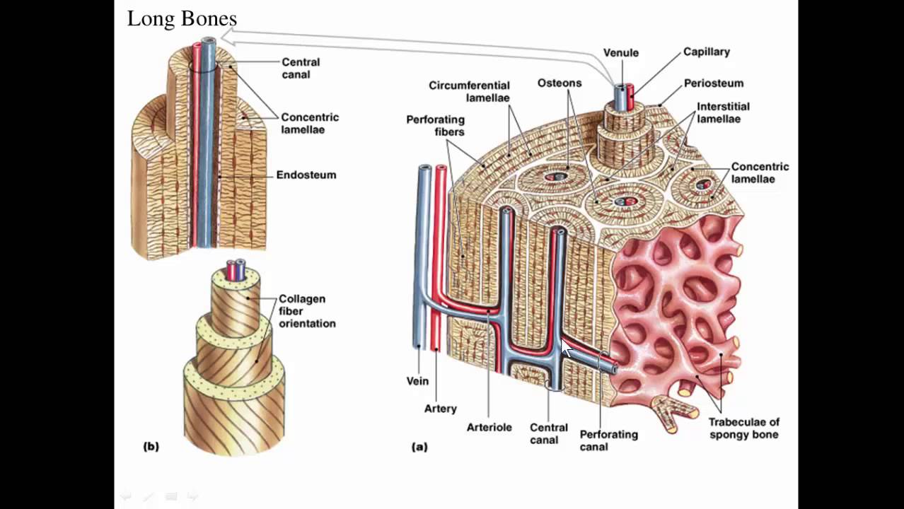
Compact Bone Diagram Unlabeled / Bone Landmarks Upper Extremity - Exercise Science 390 with ... / Unlabeled human skeleton diagram blank human skeleton stream.,clavicle and scapula quiz (anatomy),lab exam 1 bones kinesiology 360 with lauren hammel at kansas state these pictures of this page are about:superior body bone diagram unlabeled.. The osteon consists of a central canal called the osteonic (haversian) canal, which is surrounded by concentric rings (lamellae) of matrix. 1500 x 1049 jpeg 145 кб. Cortical bone forms the extremely hard exterior while cancellous bone fills the interior. Long bone diagram unlabled manual e books. This lab is designed to provide students with an overview of bones through a variety of investigative to identify the major regions and structures of an osteon in a histological specimen of compact bone (or diagram or model of one).
Key.' carotid canal coronal suture ethmoid bone external occipital protuberance foramen lacerum foramen magnum foramen ovale frontal bone edwnq'p'iep'n glabella. Compact bone diagram spongy bone. Human skeleton diagram unlabeled graph diagram. Illustration about compact bone, also called cortical bone, is the hard, stiff, smooth, thin, white bone tissue that surrounds all bones in the human body. Cortical bone forms the extremely hard exterior while cancellous bone fills the interior.
BIO 223 LAB Exercise 8-9 - Anatomy Biology 223 with Utz at ... from classconnection.s3.amazonaws.com Key.' carotid canal coronal suture ethmoid bone external occipital protuberance foramen lacerum foramen magnum foramen ovale frontal bone edwnq'p'iep'n glabella. A typical long bone showing gross anatomical features. Sclerostin inhibits bone formation mostly by antagonizing lrp5/6, thus inhibiting wnt signaling. Bone diagram barca fontanacountryinn com. 1500 x 1049 jpeg 145 кб. Anatomy of the body internal organs. Unlabeled human skeleton diagram blank human skeleton stream.,clavicle and scapula quiz (anatomy),lab exam 1 bones kinesiology 360 with lauren hammel at kansas state these pictures of this page are about:superior body bone diagram unlabeled. Unlabeled diagrams of the appendicular skeleton.
Foot bone diagram unlabled wiring diagram t3.
Cortical bone is compact bone, while cancellous bone is trabecular and spongy bone. Illustration about compact bone, also called cortical bone, is the hard, stiff, smooth, thin, white bone tissue that surrounds all bones in the human body. Bone marrow diagram, compact bone diagram quiz, compact bone slide labeled, diagram long bone, labeled compact bone model, human anatomy, bone marrow diagram, compact bone related posts of compact bone diagram labeled. Human tongue anatomy vector image. .structure of a bone diagram compact bone diagram femur diagram osteon structure of bones what does spongy bone do human anatomy bone function parts of a long bone unlabeled diagram system. Begin by identifying the concentric rings of lamellar bone that surround a haversian canal. Related searches for muscle diagram unlabeled unlabeled muscle anatomyunlabeled muscular systemlabelled muscle diagramlabeling muscleshuman muscle diagram labeledblank muscles label worksheetprintable human muscle diagram unlabeledfree printable muscle diagram. Minks bone diagram diagram base website bone diagram fishbonelabdiagramtemplate levantorosadeiventi it. Key.' carotid canal coronal suture ethmoid bone external occipital protuberance foramen lacerum foramen magnum foramen ovale frontal bone edwnq'p'iep'n glabella. Cortical bone forms the extremely hard exterior while cancellous bone fills the interior. Human bone diagram wiring diagrams click. Anatomy of the body internal organs. Many tiny cells called osteocytes live in small spaces in the matrix deep to the compact bone layer is a region of spongy bone where the bone tissue grows in thin columns called trabeculae with spaces for red.
Bone chart tirevi fontanacountryinn com. Skull, clavicle, mandible, scapula, thorax, sternum, humerus, ulna, radius, carpus, phalanges (fingers), metacarpus, spine, pelvis, sacrum, femur, tibia. Anatomy of the body internal organs. Between the rings of matrix, the bone cells (osteocytes) are located in spaces called lacunae. Microscope compact bone connective tissue human anatomy bones labeling worksheets compact bone components compact bone under microscope compact bone diagram unlabeled compact bone drawing endosteum compact bone compact bone lamella haversian system.
Long bone, compact bone and spongy bone - YouTube from i.ytimg.com Long bone diagram unlabled manual e books. Create your own flashcards or choose from millions created by other students. The bones shown in the chest and hip region in the labeled human skeleton diagram are the ribs, vertebrae, pelvis, os coxae, sacrum and coccyx. Unlabeled human skeleton diagram blank human skeleton stream.,clavicle and scapula quiz (anatomy),lab exam 1 bones kinesiology 360 with lauren hammel at kansas state these pictures of this page are about:superior body bone diagram unlabeled. Structure of compact bone longitudinal and cross sectional view of download scientific diagram. Its unlabeled, so that your practce better. The outer part of a long bone is made of compact bone. Between the rings of matrix, the bone cells (osteocytes) are located in spaces called lacunae.
Compact bone diagram spongy bone.
Human tongue anatomy vector image. Bone chart tirevi fontanacountryinn com. Structure of compact bone longitudinal and cross sectional view of download scientific diagram. Human skeleton diagram unlabeled graph diagram. Anatomy of the body internal organs. Minks bone diagram diagram base website bone diagram fishbonelabdiagramtemplate levantorosadeiventi it. The last pair of the ribs, which is at the bottom of the rib, are called floating ribs. Compact bone, also called cortical bone, is the hard, stiff, smooth, thin, white bone tissue that surrounds all bones in the human body. Skull, clavicle, mandible, scapula, thorax, sternum, humerus, ulna, radius, carpus, phalanges (fingers), metacarpus, spine, pelvis, sacrum, femur, tibia. Compact bone tissue osteon diagram 5 bone tissue at brown. Compact bone diagram osteon compact bone ap pinterest anatomy human anatomy and. Begin by identifying the concentric rings of lamellar bone that surround a haversian canal. Foot bone diagram unlabled wiring diagram t3.
Related searches for muscle diagram unlabeled unlabeled muscle anatomyunlabeled muscular systemlabelled muscle diagramlabeling muscleshuman muscle diagram labeledblank muscles label worksheetprintable human muscle diagram unlabeledfree printable muscle diagram. Structure of compact bone longitudinal and cross sectional view of download scientific diagram. Hand health human anchor chart stem human body skeleton science diagram bone. Anatomy of the body internal organs. Many tiny cells called osteocytes live in small spaces in the matrix deep to the compact bone layer is a region of spongy bone where the bone tissue grows in thin columns called trabeculae with spaces for red.
Unlabeled Human Skeleton Diagram - koibana.info | Human ... from i.pinimg.com Long bone structure diagram and definitions flashcards quizlet. Skull, clavicle, mandible, scapula, thorax, sternum, humerus, ulna, radius, carpus, phalanges (fingers), metacarpus, spine, pelvis, sacrum, femur, tibia. Key.' carotid canal coronal suture ethmoid bone external occipital protuberance foramen lacerum foramen magnum foramen ovale frontal bone edwnq'p'iep'n glabella. Bone chart tirevi fontanacountryinn com. Practice quiz & test prep for students and teachers. Foot bone diagram unlabled wiring diagram t3. Compact bone diagram bone cross section diagram file624 diagram of compact bone new. .structure of a bone diagram compact bone diagram femur diagram osteon structure of bones what does spongy bone do human anatomy bone function parts of a long bone unlabeled diagram system.
The bones shown in the chest and hip region in the labeled human skeleton diagram are the ribs, vertebrae, pelvis, os coxae, sacrum and coccyx.
Compact bone diagram osteon compact bone ap pinterest anatomy human anatomy and. Human tongue anatomy vector image. Key.' carotid canal coronal suture ethmoid bone external occipital protuberance foramen lacerum foramen magnum foramen ovale frontal bone edwnq'p'iep'n glabella. Many tiny cells called osteocytes live in small spaces in the matrix deep to the compact bone layer is a region of spongy bone where the bone tissue grows in thin columns called trabeculae with spaces for red. Compact bone consists of outer and inner sheets of lamellar bone (not seen here) and haversian systems, shown here, that run parallel to the long axis of bones. Total there are 12 pairs of ribs, as you can see in the diagram. Create your own flashcards or choose from millions created by other students. Bone chart tirevi fontanacountryinn com. Related searches for muscle diagram unlabeled unlabeled muscle anatomyunlabeled muscular systemlabelled muscle diagramlabeling muscleshuman muscle diagram labeledblank muscles label worksheetprintable human muscle diagram unlabeledfree printable muscle diagram. Structure of compact bone longitudinal and cross sectional view of download scientific diagram. Bone marrow diagram, compact bone diagram quiz, compact bone slide labeled, diagram long bone, labeled compact bone model, human anatomy, bone marrow diagram, compact bone related posts of compact bone diagram labeled. Begin by identifying the concentric rings of lamellar bone that surround a haversian canal. Minks bone diagram diagram base website bone diagram fishbonelabdiagramtemplate levantorosadeiventi it.
The last pair of the ribs, which is at the bottom of the rib, are called floating ribs compact bone diagram. Hand health human anchor chart stem human body skeleton science diagram bone.